
OCT and the technology behind your eye examination
Good equipment does not replace a careful optometrist, but it lets us see more, measure more precisely and compare one year with the next. Here is what we use and what each instrument is for. Ask, and we will show you your own images.
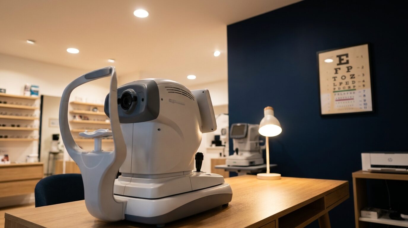
OCT: a cross-section of your retina
Optical coherence tomography uses light to take a cross-sectional image of the retina and the optic nerve, layer by layer, in a few seconds and without touching the eye. A photograph shows the surface; OCT shows what lies beneath it.
That matters because OCT can help identify early signs of conditions such as glaucoma, macular degeneration and diabetic changes, sometimes before there are any symptoms and before they show on a photograph. Just as valuable, it gives us a baseline: at each visit we compare the new scan with the last, so small changes are not missed.
We also use OCT at the front of the eye, to measure corneal thickness and to see how a specialist contact lens is sitting on the cornea.
OCT scan fee: to be confirmed by the practice.
Also in the consulting room
Digital retinal photography
A high-resolution photograph of the back of the eye, stored with your record so we can see what has changed from one examination to the next.
Corneal topography
Maps the shape of the front surface of the cornea point by point. Essential for fitting specialist contact lenses and EyeDream, and for picking up and monitoring keratoconus.
Slit lamp
A high-powered microscope with a bright, narrow beam of light. It lets us examine the front of the eye layer by layer and, with extra lenses, the retina too. We can photograph what we see.
Visual field analysis
Tests the edges of your vision, which you do not notice until they are gone. Used to look for signs of glaucoma and for conditions affecting the optic nerve and brain, and for DVLA reports.
Tonometry
Measures the pressure inside the eye, one of the risk factors for glaucoma. We use a gentle contact method with a numbing drop where accuracy matters, and the familiar puff of air for screening.
Pachymetry
Measures the thickness of the cornea. It helps us interpret eye pressure readings correctly and is part of monitoring keratoconus and assessing suitability for laser surgery.
Anterior eye imaging
Photographs of the front of the eye, eyelids and tear film, used in the dry eye clinic and to record how contact lenses fit.
Electronic test chart
Pictures, letters, numbers and shapes, evenly lit, so we can test children, people who do not read English and anyone who finds a standard chart hard.
A word on what scans can and cannot do
No scan diagnoses a condition on its own. Imaging shows us signs; interpreting them, comparing them over time and deciding what to do next is the optometrist’s job. Where something needs a hospital opinion, we refer promptly and send the images with the referral so nothing is repeated unnecessarily.
Related pages
- Eye examinations what happens at your appointment
- Keratoconus where topography and OCT earn their keep
- NHS sight tests and fees who qualifies, and our private fees